In a shocking medical case, doctors in Shanghai, China, discovered a fetus growing inside the skull of a one-year-old girl. This rare condition, known as fetus in fetu (FIF), presented severe developmental issues for the child, who experienced significant delays in motor skills and speech development.1
Initial Symptoms and Diagnosis

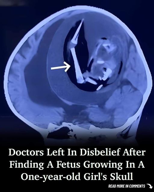
The parents of the one-year-old girl noticed her developmental delays and brought her to the hospital. Doctors performed a head CT scan, which revealed a large mass in her cerebral hemisphere. This mass, measuring 13 centimeters in diameter, contained internal bone structures and had a smooth boundary.
Prenatal Abnormalities

Abnormalities were detected during a 33-week prenatal examination, but an MRI could not provide detailed information due to space occupation in the skull. The girl was delivered via cesarean section at 37 weeks, already showing signs of a large head circumference.2
Understanding Fetus in Fetu

Fetus in fetu is a rare developmental anomaly where a malformed fetus is found within the body of its twin. It occurs in approximately one in half a million live births. Typically, FIF is located in the abdomen, but in this case, it was found in the skull.3 “FIFs remain a mystery,” said study authors Xuewei Qin and Xuanling Chen from Peking University International Hospital, highlighting possible environmental and genetic factors.
Surgical Intervention

Given the severe condition, doctors decided to perform a craniotomy, a surgical procedure that involves removing part of the skull to expose the brain. During the surgery, they discovered a malformed fetus with visible limbs, mouth, eyes, and other organs. “Following complete mass resection, mouth, eye, arm, and hand shapes could be observed,” the study reported.
Challenges in Surgery

Despite extensive preoperative examinations and planning, the one-year-old girl experienced uncontrollable seizures post-surgery. She remained unconscious and sadly passed away 12 days after the procedure. The prognosis for such cases is generally poor, with surgical resection being the only curative treatment.
Implications and Research

The rarity of intracranial FIF cases underscores the need for early diagnosis and treatment. Differentiating between FIF and other conditions like teratomas is crucial for effective medical intervention. “Monitoring alpha-fetoprotein levels after surgery can help detect recurrence,” researchers noted.
Case Study Reflections

This case adds to the less than 200 reported instances of FIF in medical literature. It highlights the importance of advanced imaging techniques and prenatal care in detecting and managing such rare conditions. The doctors’ experience in Shanghai offers valuable insights into the complexities of diagnosing and treating FIF.
Conclusion

The discovery of a fetus growing in an infant’s skull is a profound reminder of the complexities and mysteries of human development. This case emphasizes the need for continuous research and advanced medical practices to manage such rare and challenging conditions effectively.